November 8 marks the annual World Radiography Day. The Imaging team at North Bristol NHS Trust would like to take this opportunity to introduce themselves and share with you some examples of the work they do, and identify the vital contribution the Radiography profession makes to modern healthcare.
It is likely that Radiography will be involved in your care should you need to visit hospital. The profession is diverse, and is always looking at encouraging new people to join the team that is at the heart of effective and modern patient care.
Below you can read a profiles from some of our staff working in the various disciplines of Radiography.
X-Ray
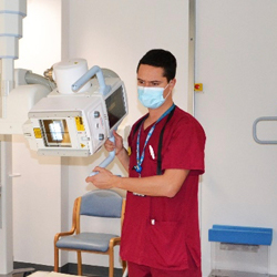
Hello my name is Liam.
I am a Diagnostic Radiographer at North Bristol NHS Trust. At this Trust we have radiographers trained in a wide range of specialities, and they all play key roles in the effective delivery of healthcare.
After completing a 3-year degree with academic study and clinical practice, every radiographer starts off like me, in plain film x-ray. As a Plain Imaging radiographer I perform diagnostic X-Ray Imaging in a variety of clinical settings. This includes inpatient X-Ray for those patients staying in the hospital, outpatient X-Ray for patients that are visiting clinics and referred from their GPs, Imaging for A&E patients, X-Ray imaging for patients receiving surgical theatre procedures, and ward based mobile imaging for those patients too unwell to travel to our department. Every day is different, every patient is different, and it is vital we maintain a flexible approach so that we can deliver high quality imaging, often in challenging circumstances, throughout the 24 hour day.
2020 has been a tough year for everyone working in the hospital due to Covid-19, and x-ray and CT have been key in the treatment of patients with this disease. Similarly to other hospital workers, we spend hours in PPE, protecting ourselves and the patients we treat.
The majority of patients that enter our hospital doors will encounter a radiographer at some point. At North Bristol NHS Trust, radiographers perform on average over 1000 patient examinations a day. World Radiography Day allows us to showcase our fantastic staff who work tirelessly across a wide range of different modalities.
Our list of work is endless but hugely fulfilling and can be seen to benefit every patient we see.
Sonography
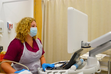
My name is Grace and I started my ultrasound training 3 years ago following working within plain film Radiography for 2 years. Since then I have completed a post-graduate diploma in Medical Ultrasound. Ultrasound uses sound waves to create images of the inside of the body; it is especially good at imaging solid organs and soft tissues. Personally, I am trained to scan and report abdomens, the female pelvis, testes and thyroids. These reports are communicated with the referring doctor to guide a patient’s clinical management.
An example of a common reason for performing an ultrasound of the abdomen would be to assess for gallstones and associated complications such as gallbladder infection or blockage of the biliary system.
As sonographers we have a very varied role and work in a large number of clinical settings; within the Emergency department or emergency clinics such as Surgical Hot Clinic, Urology clinic, Emergency Gynaecology clinic and the Early Pregnancy Clinic. We also scan inpatients either in the imaging department or portably on the wards and perform routine and surveillance outpatient scans at Southmead and within the community sites.
MRI
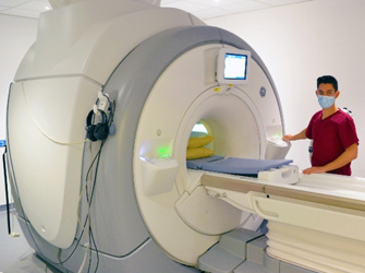
My name is Alex and I’m an MRI Radiographer
MRI radiography has revolutionised the way doctors look at cancer, brains, spines and muscles. We make high definition images of the human body using magnets that have the strength to pick up a car. Should the patient have any concerns about lying down in the scanner for their examination we can help assure them and make them as comfortable as possible so that they can receive the best Imaging possible. Working alongside the other imaging areas, our images help doctors decide what drugs and surgery patients will have. As a regional centre for neuroscience and the skeleton, patients travel from as far as Sussex, Cornwall and even Ireland to get specialist scans done here.
At Southmead Hospital we are one of the largest MRI departments, with 4 scanners worth about £2 million each. After finishing a degree in radiography, and often a period working in x-ray or CT, you can specialise in MRI. Many people in our department have been sponsored to study a masters-level MRI qualification at university.
I love working as a radiographer because I get to meet all kinds of different people that come to us with different problems. I chose to specialise in MRI because the images of the human body are so detailed and I am also fascinated with state of the art technology.
CT
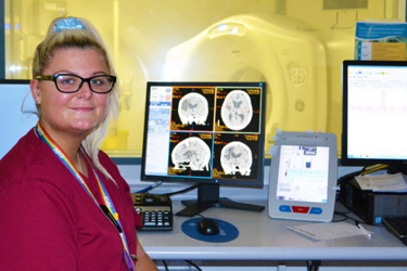
My name is Holly, and I am a CT Radiographer.
CT or Computed Tomography uses X-rays to create cross sectional images. This diagnostic tool can be used for diagnosing cancer, fractures, internal bleeds, and cardiovascular diseases and even used in guided needle biopsies, amongst many other things. I had worked in plain film x-ray for a year before specialising in Computer Tomography. Since spending the past year in CT I have been able to further my skills in decision making and working under pressure. I work closely with the Emergency team to prioritise patients who are in need of rapid diagnoses of life threatening injuries, including major trauma, acute strokes and critical pathologies.
These services run 24/7 to optimise patient care. Another area in the hospital where CT is used is in the primary role of diagnosis and staging of many cancers, while also using later scans for follow up assessment of the disease progression. CT is widely used in hospitals for quick diagnosis of patient’s conditions, it is vital in delivering accurate and timely diagnostic Imaging for effective decision making to benefit the healthcare outcome for the patient.
Nuclear Medicine
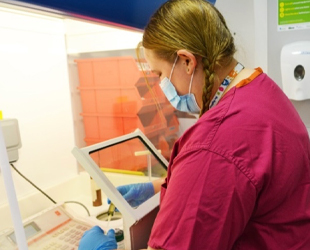
My name is Vicki and I am a diagnostic radiographer in my second year of training as a Nuclear medicine practitioner. To become a nuclear medicine practitioner you would first complete an undergraduate degree in diagnostic imaging or equivalent, and then go on to a post graduate qualification in Nuclear medicine. A Nuclear medicine practitioner is defined as either a radiographer or technologist who will prepare radioactive pharmaceuticals and administer them to patients for imaging or therapeutic purposes.
A typical day in department for me would include explaining radiographic procedures to the patient and answer any questions they may have. I prepare radioactive pharmaceuticals and administer them to the patient by way of injection, eating of a meal or in pill form. I operate imaging equipment which includes both nuclear medicine emission scanning and computed tomography (CT).
We interact with many healthcare professionals in our role including Cardiology, Plastic surgery, Urology, Orthopaedics and Neurology covering a range of scans like kidney renograms, brain DAT scans, cardiac stress scans, bone scans and breast and melanoma sentinel node localisation. Whereas other modalities look at the anatomy of the human body, as nuclear medicine practitioners we look at function and physiology of the human body.
Reporting
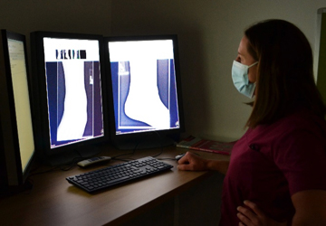
My name is Laura; I have been a diagnostic radiographer for 11 years where I have been fortunate enough to expand my professional skills. I have just recently achieved my current role as a Musculoskeletal reporting radiographer by completing a two year post graduate diploma from Exeter University in Medical Image interpretation.
It has been recognised that radiographers are well placed to be upskilled into advanced practise roles such as plain film image interpretation. There is an ongoing national shortage of radiologists who are the doctors responsible for the interpretation of diagnostic images taken by radiographers. This shortage has seen an increase in diagnosis waiting times for patients and therefore upskilling radiographers into these roles can improve patient pathway.
My role is to interpret musculoskeletal plain films from a range of referral sources such as GP, ED, orthopaedics and rheumatology. The interpretation and communication of a plain film is a key element in ensuring the right pathway for the patient. For example, a GP patient is referred for an x-ray and an abnormality is demonstrated, my role is to ensure I convey clearly the next best step for that patient.
Reporting is a challenging role where a vast breadth of knowledge is required to produce accurate and timely reports. However the additional responsibility gives you a great increase in personal and professional development and a sense of achievement.
Mammography
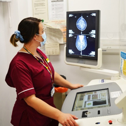
Hello, my name is Mary. I am the Deputy Superintendent Radiographer for Avon Breast Screening, Breast Care Centre. I have been a Radiographer for 7 years. I did 2 years post qualification as a Plain film Radiographer in the ‘old’ Southmead Hospital and helped in the move to the ‘new’ Brunel Building.
The training to become a Mammographer is roughly a year at master’s level, and results in attainment of a Post Graduate certificate in Mammography. The training primarily requires trainees to keep a log book and obtain around 500 mammograms, which you are appraised on with an image assessment via face to face tutorials with a qualified trainer, plus various essays and case studies.
Although Mammograms are our bread and butter, we also train to do further imaging techniques, such as Tomosynthesis (3D imaging), Magnification views, Wire localisations and Stereotactic Core Biopsies. We work very closely with Radiologists, Assistant Practitioners, Nurses, Radiography Assistants and Admin and Clerical staff who all work fantastically together – one of the advantages of being in a smaller department is that it is very close knit and staff are encouraging and supportive of each other.
We are one of the largest Breast Centres in the UK, running a Symptomatic ‘one-stop’ service for women (and men!) who have been referred via their GP, and we provide a National Breast Screening Programme to women aged 50-70 across Bristol and surrounding areas. We do approximately 60,000 Mammograms per year which keep us very busy!
We have two static sites, Southmead Hospital and Central Health Clinic in the city centre where we do a majority of our screening and two mobile units that are fully Radiographer lead, serving the communities surrounding Bristol e.g. Shepton Mallet, Weston-Super-Mare, Portishead, Bath, Clevedon, Thornbury and Paulton.
Our job is varied and fast paced, and each day presents with new challenges. We have to be flexible to ensure we are providing the best service to our clients and patients. Mammographers have to work quickly and efficiently in order to provide high quality diagnostic images whilst being professional and reassuring clients who are anxious, scared and quite often in pain.
Whilst we pride ourselves in providing excellent patient cantered care, we also value the need for skill mix and other opportunities for staff such as Quality Assurance, EROS and Audits and Advance Practice – we are also fortunate enough to have Radiographers here who have advanced to Consultant Radiographers.
Interventional
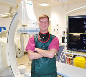
My name is Matt and I’m a radiographer based in Interventional Radiology. I qualified 5 years ago and spent the vast majority of that time in Interventional Radiography (IR) after initially starting in plain film x-ray.
As an IR radiographer, it is our responsibility to control state of the art equipment and make sure our radiation dose is as low as reasonably practicable whilst the procedure is ongoing. We work as part of a multidisciplinary team involving radiologists, surgeons, nurses, HCA’s and so effective communication between all staff is crucial.
Most IR procedures are minimally invasive alternatives to open or laparoscopic (keyhole) surgery which is always a bonus to patients if that option is available. These procedures consist of removing a blood clot causing a stroke, inserting drains into septic collections or to stop life-threatening bleeding caused by a variety of conditions. Part of our scope even includes cardiac scanning, looking at vessels in the heart. Being the regional vascular, neuro, urology and trauma centre, NBT covers a wide area ranging from South Wales, Wiltshire, Devon and Gloucester which is great as you get to meet all different kinds of people and be a part of their recovery process.
Fluoroscopy
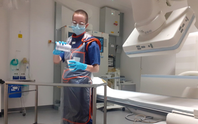
My name is George, I am a diagnostic radiographer specialising in gastro-intestinal fluoroscopy. The use of “live” x-rays allows the function of the body to be investigated, as well as the anatomy. I am trained to carry out and report on barium swallow examinations, which are used to investigate swallowing difficulties, cancers, hernias, reflux, and obstructions for example. I also do some procedures to rule out bowel leaks following surgery, and to examine pelvic floor function.
It is a varied day typically involving a wide variety of procedures. A GI specialist radiographer will usually lead their own lists, but we also facilitate other procedures which are led by radiology doctors or other specialist radiographers.
Fluoroscopy plays a huge role within the hospital for both inpatients and outpatients, and I am proud of the flexibility our service offers.
